Surgical Endoscopy

About Surgical Endoscopy

Surgical Endoscopy is an umbrella term describing surgery using small rigid endoscopes through small (‘keyhole’) incisions. Surgical Endoscopy includes laparoscopy (single to multiple incisions into the abdomen), thoracoscopy (single to multiple incisions into the chest cavity) and ‘NOTES’ (Natural Orifice Transluminal Endoscopic Surgery aka ‘scarless surgery’ using natural holes without the need of external incisions). 
 
At EVR, we only perform laparoscopic procedures in the general practice setting. More advanced endoscopic surgical procedures are considered on case by case basis and performed at a multi-disciplinary tertiary hospital. 

Our Procedures

Laparoscopy is a procedure that allows the veterinary surgeon to directly examine the abdominal cavity and organs, as well as to undertake minimally invasive surgical interventions (keyhole surgery). A laparoscope is a small rigid endoscope with a camera and a light source. As in human surgery, in order to perform laparoscopy, a working space needs to be created within the abdomen. This is achieved by inserting a needle into the abdominal cavity and inflating it with carbon dioxide. Once the abdomen has been inflated, a cannula (small plastic or metal tube) can be inserted through a small (0.5–1 cm) incision in the skin and muscle of the abdominal wall and directed into the abdominal cavity. The laparoscope is placed through this cannula. Once the laparoscope is in place, the remaining cannulae for the surgical instruments can be placed. A typical laparoscopic procedure may require the placement of between one and four cannulae.

Laparoscopy may be recommended for diagnostic and/or interventional (treatment) purposes.

Minimally invasive therapeutics:

A variety of other procedures can be performed with little or no discomfort to the patient. The endoscopist can remove the ovaries (ovariectomy or ‘lap spay’), the ovaries and uterus (ovariohysterectomy), retained abdominal testicles (cryptorchidectomy) and the gall bladder (cholecystectomy), perform a gastropexy (suture the stomach to the abdominal wall to reduce the risk of gastric torsion in predisposed breeds), remove bladder stones, remove foreign bodies and remove tumours (e.g. in the adrenal glands or spleen).

Less pain is provided with laparoscopic spays due to small incision size(s), decreased muscular trauma and the relatively atraumatic nature of cauterisation and transection of the ovarian pedicle compared with digital disruption of suspensory ligament during traditional midline ovariectomy. A further benefit of laparoscopic surgery is the superior visibility of internal structures compared with open procedures. Small incisions are often the pride of many vets performing traditional midline OVE or OHE; however, this method results in decreased visualisation of the reproductive tract. This can result in both pain from traction of the ligament and also result in a high risk of incomplete tissue resection. This can cause ovarian remnant syndrome.
 
 
Single incision Ovariectomy (‘Lap Spay’):
 
 
EVR can perform single incision ovariectomies, via the umbilicus (‘belly button’), using a special operating telescope. Although you may have heard of vets doing a ’normal spay’ though a tiny incision, the difference between the two is what happens on the inside! 
 
Non-laparoscopic spays, unlike laparoscopic spays, rely on releasing the ovaries from their attachment site (please read EVR/BSAVA Client Information Sheet on Laparoscopic Spays). To achieve this, the ligament needs to be blindly and manually torn away from the peritoneum using blind digital traction. After releasing the ovary from it’s attachment site, it’s then pulled towards the tiny incision for its removal. This technique not only results in a tremendous amount of pain for the pet, but it also risks leaving behind pieces of ovary within the abdomen. Retained ovarian tissue, regardless if performing an ovariectomy (removing ovaries only) or ovariohysterectomy (removing ovaries and uterus), will result in a repeated heat cycles (ovarian remnant syndrome). 
 
A single port laparoscopic spay benefits from 1) a single 1-1.5 cm incision (dependant on size of ovary to exteriorise) in the belly button and 2) the gentle suspension and sealing of vessels which release it from its attachment site. This is all under direct visual guidance which ensures the entire ovary has been removed. This method eliminates the technique of blindly tearing the ligament from the abdominal wall and the risk of leaving ovary remnants behind whilst trying exteriorise through a tiny incision. 
 
Increased duration of surgical times have been reported to be a negative factor in laparoscopic surgery; however, surgical times do depend on surgeon’s level of experience, instrument availability and the kind of procedure being performed (ovariectomy or ovariohysterctomy). 
 
At EVR, our surgical times for both single and two-port ovariectomies (from initial incision to closure) are anywhere between 10-12 minutes. For larger and more active dogs, it may be advantageous to consider a single port approach due to fewer wounds and risk of multiple wound breakdown.
 
Two-Port Ovariectomy and Ovariohysterectomy:
 
This method is similar to Single Port Laparoscopic Ovariectomy; however, two access ports are used to remove the ovaries +/- uterus. A two port and three port procedure is considered more routine in the UK.
This is a procedure which permanently fixes the stomach to the body wall to help prevent gastric dilatation volvulus (GDV) in at-risk breeds. This can be achieved using one of two techniques 1) Laparoscopic-assisted Gastropexy or 2) Total Intracoporeal Gastropexy. A laparoscopic-assisted technique involves suturing the stomach, via an external incision along the side of the body wall, to fix the stomach in it’s new position. 
 
A total intracorporeal gastropexy is a technique of using barbed suture to permanently fix the stomach to the abdominal wound without the need for an  additional external incision. The benefits of total intracorporeal gastropexy is reducing the risk of external wound swelling, pain and wound breakdown. At EVR, we perform both laparoscopic-assisted and tota invasive technique intracorporeal preventative gastropexies. 

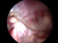
This is a minimally-invasive technique used to not only diagnose the location of retained testicle(s) (i.e. inguinal canal vs intra-abdominal) but also assists in the removal of intra-abdominal testicles through a single access port (SILS Port) via the umbilicus (belly button). 

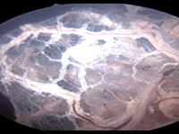
This is a minimally-invasive technique that allows for the full exploration of the bladder, trigone, ureteric openings and proximal urethra via a small abdominal incision and tiny bladder incision for the cystoscope (rigid endoscope).  This also allows for interventional treatment(s) including visual removal of bladder stones using basket retrievers/graspers, laser ablation of bladder polyps and laser ablation of transitional cell carcinoma. The procedure uses continuous saline lavage which not only enhances visualisation but also useful for removing retained urinary debris which can be irritate bladder lining.

This is a minimally invasive technique which allows for a full laparoscopic exploration of the abdominal cavity whilst also allowing for complete removal using vessel sealer technology and a wound retractor device. This results in a faster procedure and smaller abdominal incision compared to traditional open surgery*. 
 
*NOTE: This procedure is only performed in suitable cases/patients and highly dependant on spleen size and cause of underlying condition requiring splenectomy.
This is an advanced laparoscopic procedure to remove the gallbladder.  Case selection is paramount and not all patients with gallbladder mucocoele are candidates for keyhole surgery. Emergency cases, cases with concern about impending gallbladder rupture and wall compromise as well as patients with significant increase of bilirubin and biliary duct distension/obstruction (who may need duct flushing via open surgery) are not suitable candidates. Suitable candidates for laparoscopic cholecystectomy are cases which are considered elective, have early gallbladder mucocoele and no elevation of Tbil.
 
*NOTE: This procedure is only performed in a tertiary multidisciplinary specialist hospital setting.

We offer an experienced, accessible, approachable, reliable and time-efficient suite of endoscopy services for veterinary colleagues based in general practice settings, secondary clinical centres and tertiary multidisciplinary specialist hospitals.