Lower GI endoscopy, often referred to as ‘ileoscopy and colonoscopy’, is a procedure that allows the veterinary surgeon to directly examine the lining of the lower part of the GI tract. The lower GI tract consists of the last part of the small intestine (distal jejunum/ileum) and the colon, including the caecocolic pouch (similar to the appendix in humans) and the rectum.
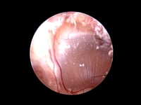
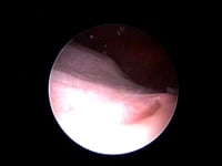
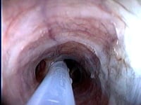
A flexible tube with a camera and a light source, about the thickness of your little finger, is placed into your pet’s rectum to examine the last part of the colon (rectum and descending colon), the middle colon (transverse colon), the first part of the colon (ascending colon) and the ileocolic sphincter (the valve that separates the last part of the small intestine from the colon).
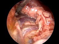
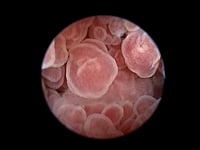
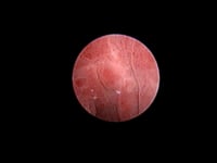
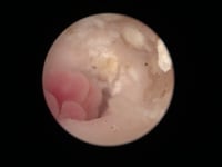
Colonoscopy and ileoscopy are performed to evaluate the signs and attempt to identify/exclude the causes of persistent diarrhoea, weight loss, low vitamin B12 levels, excessive mucus, straining to defaecate and blood in the faeces (haematochezia). It is an excellent minimally invasive alternative to traditional surgery and tends to be more accurate than abdominal imaging (radiography and ultrasonography) for detecting inflammation (redness), bleeding, ulcers, polyps and tumours.
Minimally invasive diagnostics:
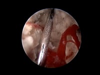
Endoscopy can be used to obtain biopsy samples using specialised instruments known as biopsy forceps. Biopsy samples are routinely obtained during endoscopic examinations as many conditions can only be diagnosed in a laboratory by analysing tissue specimens. Biopsy samples from suspicious areas can be useful for distinguishing between cancerous and non-cancerous conditions.