Interventional Endoscopy + Endoscopic Laser

About Interventional Endoscopy

Interventional Endoscopy is a term which describes a broad range of procedures which can be performed during an endoscopic examination. These can include foreign body removals, polypectomy, gastric feeding tube placement, nasal/sinus fungal debridement and anti-fugal soaks, laser and balloon dilatation of strictures, laser tumour debulking, laser ectopic ureters.

Interventional Endoscopic Procedures

Minimally invasive interventions:

A variety of instruments can be passed through the endoscope, allowing the veterinary endoscopist to treat a range of abnormalities with little or no discomfort to the patient. The endoscopist can remove swallowed objects (foreign bodies) using grasping forceps, baskets or snares, stretch narrowed areas (strictures) with a balloon catheter, remove polyps or treat upper GI bleeding. Endoscopy can also be used for the placement of feeding tubes that bypass the oesophagus and open directly into the stomach.

Minimally invasive interventions:

A variety of instruments can be passed through the endoscope, allowing the veterinary endoscopist to treat a range of abnormalities with little or no discomfort to the patient. The endoscopist can stretch narrowed areas (strictures) with a balloon catheter, remove polyps, treat (‘debulk’) tumours or even treat lower GI bleeding using a laser.

Minimally invasive interventions:

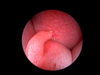
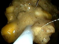
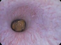
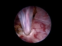
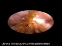
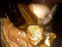
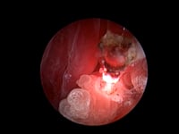
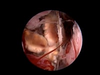
A variety of instruments can be passed through the rhinoscope, allowing the veterinary endoscopist to treat a range of abnormalities with little or no discomfort to the patient. The endoscopist can remove nasal foreign bodies (grass seeds, sticks), grass blades that have been vomited up over the soft palate in cats, fungal plaques (aspergillosis) and polyps, debulk tumours using special instruments placed through the operating port of the endoscope (e.g. lasers), debride dead turbinate tissue in cats with herpesvirus-induced rhinitis, stretch narrow areas at the back of the nose (nasopharyngeal stenosis) induced by chronic inflammatory conditions using a balloon catheter and/or laser, and use the fluid port on the endoscope for nasal/sinus flushing when continuous saline irrigation or antifungal treatment for aspergillosis is required.

Minimally invasive interventions:

A variety of instruments can be passed through the endoscope, allowing the veterinary endoscopist to treat a range of abnormalities with little or no discomfort to the patient. The endoscopist can remove inhaled objects (foreign bodies) using grasping forceps, baskets or snares, stretch narrowed areas (strictures) with a balloon catheter, remove polyps and treat airway obstructions.

Minimally invasive interventions:

A variety of instruments can be passed through the endoscope, allowing the veterinary endoscopist to treat a range of abnormalities with little or no discomfort to the patient. The endoscopist can remove foreign bodies (e.g. grass awns) using grasping forceps, remove polyps or abnormal growths using snares or a surgical laser, treat cholesteatoma and instil depot medications deep in the ear to treat resistant infections (e.g. Pseudomonas).

Minimally invasive interventions:

A variety of instruments can be passed through the endoscope, allowing the veterinary endoscopist to treat a range of abnormalities with little or no discomfort to the patient. The endoscopist can remove bladder stones and foreign bodies (e.g. grass awns) using grasping forceps, baskets or snares, stretch narrowed areas (strictures) with a balloon catheter, remove polyps, debulk tumours using a laser, treat ectopic ureters with laser ablation and instil bulking agents into the urethra to ‘tighten’ the sphincter and help reduce sphincter mechanism incontinence. Laparoscopic cystoscopy may be performed as a minimally invasive alternative to open surgery in those cases where access to the bladder via the urethra is not possible (e.g. male cats) or where large stones need to be removed.

Endoscopic Laser Surgery

The marriage of endoscopy and surgical lasers has offered veterinary endoscopists the opportunity to perform minimally invasive interventions in circumstances which were traditionally treated by more invasive surgical approaches.

At EVR, we use an 810nm:980nm dual-wavelength diode laser which provides us with the advantages of both wavelengths (vessel ablation and tissue debulking) whilst reducing risk of collateral tissue damage due to thermal spread.

Endoscopic Diode Laser Procedures

Respiratory Tract

The use of lasers, together with endoscopy, allow for a range of respiratory conditions to be treated in a curative, or palliative manner. Laser endoscopy is indicated for congenital conditions, tumours, as well as vaporisation of polyps, stenosis (nasopharyngeal, laryngeal, tracheal) and granulomas.

Primary intranasal tumours represent approximately 1% of all neoplasms in dogs and cats (mainly carcinomas, sarcomas and lymphoma). They have progressive local invasion and a low metastatic rate. It is for this reason, and because of the high recurrence rate, that local treatment has great importance. Radiotherapy is the treatment of choice, but chemotherapy or non-steroidal anti-inflammatory drugs (NSAIDs), or a combination, can be used with differing degrees of effectiveness. It is unclear whether adjuvant cytoreduction (laser rhinoscopy or open surgery) decreases recurrence or increases median survival time. Empirically, local clinical signs appear decreased, especially if access to radiotherapy is not possible. With laser turbinectomy, there is an evident reduction in the clinical signs associated with nasal masses (e.g. epistaxis, mucus and sneezing) which can buy relatively good quality of life for those patients where radiotherapy and/or chemotherapy have been declined or not available to the patient. The main palliative benefit is through increasing the airway space for improved breathing for the patient.

The procedure of choice for nasopharyngeal stenosis is balloon dilation and nasopharyngeal stent placement. However, in cases where the stenosis is located in a caudal position, and the stenotic wall is very thin and therefor the use of laser endoscopy may be sufficient alone. In cats especially, making 2 lateral cuts fron the stenotic opening allows a more controlled balloon dilation and preserves the edges of the wound to allow epithelialisation. The laser can also be used to control haemorrhage post dilation. Stents in cats are less successful as they tend to get hair wrapped around them.

Canine sinonasal aspergillosis is a relatively common disease in dogs. The main clinical signs are a profuse mucoid to haemorrhagic chronic nasal discharge, epistaxis and ulceration of the external nares with crusting. Diagnostic imaging (computed tomography (CT) and rhinoscopy) is mandatory to locate the fungal plaques, assess the sinuses and the nasal turbinate destruction, and in severe cases the cribriform plate destruction.

Treatment involves using a diode or holmium:YAG laser to ‘burn’ the fungal plaques and perform a partial turbinectomy around them.  Following the surgery, the nasal cavity is then completely filled with 1% clotrimazole cream. This is achieved by instilling the clotrimazole cream via a catheter that is inserted either through the working channel of the endoscope.

Urinary Tract

Ectopic ureters are classified as either intramural (tunnel through the caudal bladder; approximately 85% of ectopic ureters in dogs) or extramural (do not tunnel through the caudal bladder; typical in cats). This distinction allows the practitioner to select therapies best suited for the patient. Surgical correction methods are available for both classifications; however, only ectopic ureters travelling intramurally can be managed using diode laser ablation. Cystoscopy usually the easiest and the most accurate method of diagnosis of ectopic ureters. However, fluoroscopic-aided contrast urography, CT with contrast urography, or cystoscopic-assisted retrograde contrast ureteropyleography with fluoroscopy permit identification of a transmural orientation of intramural ectopic ureters.

Vestibulovaginal septal remnants can distort or block the vaginal and urethral openings. Their clinical significance is variable, but some are associated with urinary tract infection or interference with parturition. Most remnants are narrow and easily broken digitally. Those that are more difficult to detach can be freed from their dorsal and ventral attachments using a laser.

We offer an experienced, accessible, approachable, reliable and time-efficient suite of endoscopy services for veterinary colleagues based in general practice settings, secondary clinical centres and tertiary multidisciplinary specialist hospitals.